aortic aneurysms, and peripheral aneurysms.
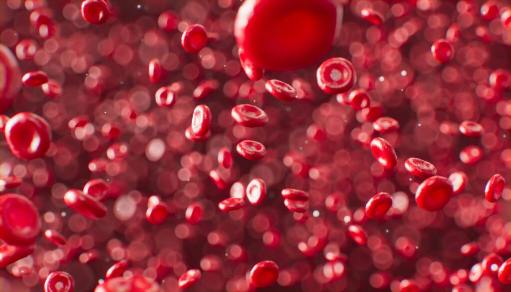
The biggest risk linked to it is a possible rupture and, thus, the ensuing life-threatening internal bleeding. Also, an aneurysm can cause other complications by way of pressing on or obstructing blood flow. There might be cases when someone gets the sickness and not knowing it. Many aneurysms do not manifest symptoms until they swell or burst.
The number of this condtition cases is quite numerous. The estimation of their actual prevalence is unattainable. It is due to the hidden character of numerous cases throughout the period of a person's life. According to recent studies, if American people were examined, it is the consensus that only 1-2% of the population might be found with a cerebral type, which would otherwise not have been detected. It is said that abdominal aortic variants are found in about 8 out of a hundred men aged over 60 and not more than 2 out of a hundred women of the same age.
Among children, it can be diagnosed while they are still very young. In U.S. adults, however, a higher number suffer from aneurysms. However, this happens as they progress in life and not during their growth phase only. In addition, the incidence is directly related to the age of the person. It is especially high in the presence of risk factors such as hypertension and active smoking.
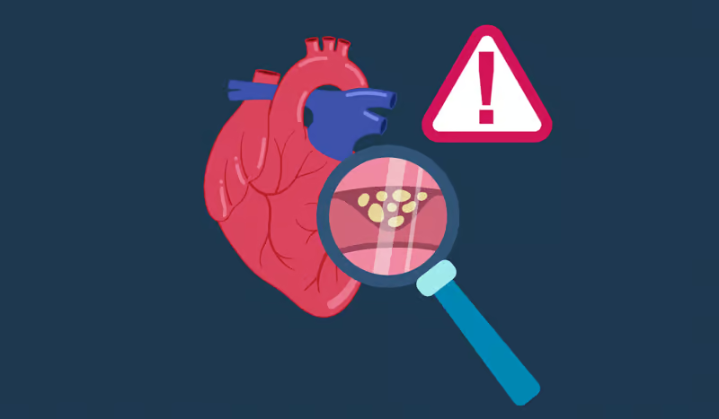
The origin of aneurysms can be due to a multitude of factors, many of which are complex and multifactorial. Factors that contribute to the development of them: genetic, environmental, or combined. This statement suggests that hypertension represents the main cause of the stiffening of the walls of arteries. At the same time, the situation is similar, where fatty materials and atherosclerosis accumulate in the vessels and become weakened. As a result, it leads to the formation of aneurysms.
Presently, as per various research studies, it has been conclusively proven that people affected by these diseases have a high probability of being affected by aneurysms. Research conducted today has evolved to the point where it is feasible to identify cases of unruptured aneurysms during cancer scans inadvertently.
On the other hand, causes related to genetics and certain heritable conditions, such as Marfan syndrome, Ehlers-Danlos syndrome, and polycystic kidney disease, can predispose an individual to aneurysms. Thus, another thing to be kept in mind is the potential infection of the artery walls or so-called mycotic aneurysms by not a large, but still a relevant number of cases.
Traumas and such, like the harm caused by physical injuries, can result in disorders in the blood vessels, leading to aneurysms either directly or via other indirectly connected routes. In addition to this, very detrimental are also the decisions made by each person regarding their lifestyle, which has a direct relationship to increased risk through smoking and consumption of alcoholic beverages upwards.
Most small and unruptured aneurysms do not cause any symptoms and may remain unnoticed for a long time. However, large aneurysms can put pressure on tissues and nerves. This causes a person to develop several symptoms according to the site of aneurysms.
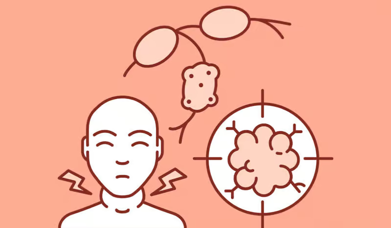
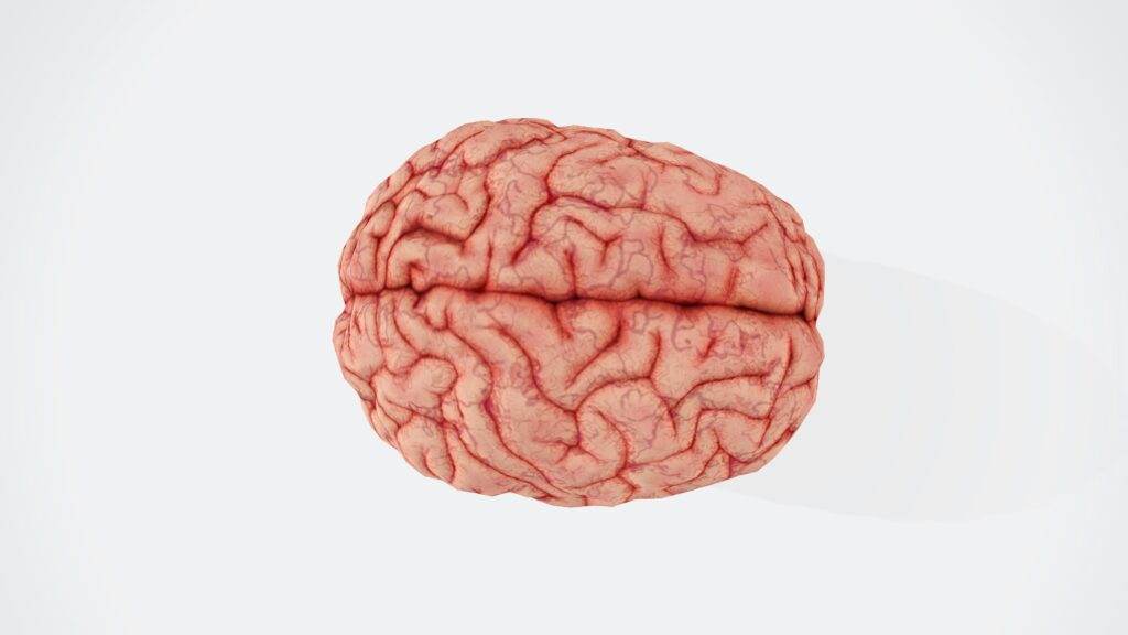
Brain aneurysm symptoms usually come in the form of a sudden and very severe migraine, loss of sight, pain near or around the eye, trouble talking, one hand or a foot, which is painless or weak, and seizures. What is heard mostly is that ruptured cerebral aneurysms have the characteristic symptoms of a sudden, very severe headache that even is commonly called the “worst headache of my life” at the same time that nausea, vomiting, stiff neck, and loss of consciousness occur.
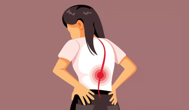
Stomach aortic aneurysms may bring about the feelings of a strong and regular pulsating by the navel. It is accompanied by dull pain in the abdomen or side and back pain. Chest aortic aneurysms are usually the cause of chest pain, cough, hoarseness, and breathing difficulties.
The symptoms assigned to this condition may be very general as well as hardly recognizable. That is the reason why diagnosis is only possible using imaging studies. With regard to this, imaging is a very important factor.
The following methods are applied to identify Aneurysm:
The confirmation of aneurysms usually comes through imaging with which the doctor is able to trace the blood vessels and pinpoint any anomalies. Computerized Tomography (CT) scans are highly effective. Through these scans, the doctor can quickly identify and diagnose bleeding in the brain and the patient might take a CT scan as the first test if he/she thinks there has been a brain type rupture.
A Magnetic Resonance Imaging (MRI) scan is another powerful weapon in the doctor's arsenal of detection. An MRI offers precise visual images of blood vessels and, thus, it is well suited for the detection of unruptured aneurysms. Additionally, Magnetic Resonance Angiography (MRA), a modality of MRI, particularly focuses on the arteries for detecting and sizing them.
Cerebral angiography continues to remain the modus operandi in the detailed diagnosis of cerebral aneurysms. In this procedure, a catheter is inserted into an artery from a femoral or brachial access and moved to the brain. A contrast agent is injected to visualize the blood vessels on X-ray images. It is a more invasive method compared to CT or MRI, but it provides highly detailed images that even allow the discovery of small aneurysms.
Ultrasound scanning is the primary screening technique for the detection of abdominal aortic types. A standard abdominal ultrasound test is a non-invasive, low-cost, and widely available method that helps confirm the diagnosis and, at the same time, signifies the size of the condition, which is important for periodic follow-up tests.
CT Angiography technology records the CT images acquired with contrast material injections to produce extended, well-distributed vascular images that are suitable for the identification of different aneurysms in the body. It is a less invasive method than conventional angiography. The resolution is high enough to be capable of displaying minute aneurysms.
Treatment strategies used to tackle Aneurysm encompass the following:
One of the classical methods of treating the brain aneurysm is to perform surgical clipping. In such a case, a small metal clip is placed at the neck of an aneurysm by a neurosurgeon. It is to prevent the flow of blood to it. This stops it from breaking or bleeding. The procedure is very invasive. That is because the skull is opened to reach the Aneurysm. However, it is known to be very effective in some cases.
Endovascular coiling is one of the major breakthroughs of the modern-day in the treatment of an aneurysm. The involved technique is minimally invasive, and a catheter moves from the blood vessels into the brain aneurysm. The micro-coils made of platinum are placed directly into the Aneurysm, packing the sac with a blood clot, and thus, the Aneurysm is sealed from the artery. The recovery periods after this procedure are typically shorter than for traditional surgery. However, coiling is not viable for all types of aneurysms.
Flow diverters are a new type of endovascular device. They are used in the treatment of some brain variant, which are large or wide-necked. These are devices that act like stents and are placed so that the blood flow is away from the Aneurysm, enabling it to heal itself. These diversions have grown popular as they present an opportunity for those who were considered to be untreatable or almost impossible to treat.
The traditional approach to treat the abdominal aortic version is open surgical repair. During the course of this surgery, a section of the aorta that is damaged is exchanged for a synthetic graft. Definitely effective, the surgery is known to be a major one requiring a very long recovery period. It is generally recommended only for candidates having large symptomatic aneurysms who tolerate the associated risk of surgery.
Endovascular aneurysm repair, or EVAR, is the procedure used in the treatment of abdominal aortic aneurysms instead of open surgery. In the process of EVAR, a stent graft is advanced into the Aneurysm through small incisions in the groin. The weakened artery wall is then supported by the stent graft, and the Aneurysm may be prevented from rupture. Among the most evident benefits is that one will have to stay in the hospital for a shorter period of time and recover quickly. However, long-term monitoring is necessary.
Plateauing of blood pressure is one of the major concern in the quest for treatment of patients with aneurysms, whether surgery is undertaken or not. There are drugs like beta blockers and ACE inhibitors available that help to lower the blood pressure and stress on vessel walls, which in turn, will reduce the possibility of its enlargement or rupture.
Doctors may, for small, asymptomatic aneurysms, and mostly for those found coincidentally, maintain this strategy of looking at them only. The more reliable strategy is to watch what happens with the help of regular imaging studies. Intervention becomes necessary if the Aneurysm increases its size beyond a certain point or gets bigger with symptoms arising in the patient.
Not only are aneurysms serious medical conditions due to the high potential for having severe complications, especially when the Aneurysm is ruptured, but also the most feared complication bears the name hemorrhage, which refers to the condition of the blood that results in a stroke, severe hypotension, organ malfunction, or death due to the location and the amount of blood. When a brain aneurysm bursts, as in this case, a subarachnoid hemorrhage occurs, which can result in permanent brain damage and, if left untreated, in a coma or death.
Even unruptured aneurysms can sometimes cause serious issues. They may grow large enough to press on nearby nerves, vessels, or organs. As a result, they cause pain, difficulty in movement, swallowing problems, or vision loss. Blood clots, or embolisms, can form inside some aneurysms. These clots can dislodge and travel to other parts of the body, causing strokes or ischemia.
Rupture does not always occur, and in some instances, type such as the abdominal aortic may leak progressively over a long period before the final catastrophic event of its breakdown, a condition known as a “sentinel bleed.” Infections and inflammations of the Aneurysm, conditions though unlikely, could complicate the treatment and also involve an increased risk of mortality. Moreover, the treatment process itself, especially if requiring surgery, is not without risks of complications, which include infection, bleeding, and recurrence.
They vary in terms of location and appearance, which leads to their classification into separate categories. The foremost types are the aneurysms of the brain (cerebral aneurysms), aortic aneurysms, and peripheral aneurysms.
About cerebral types, they are most likely to be found in the blood vessel that is adjacent to the brain and are thus referred to as intracranial types. They are typically located at the junction of arterial branches at the base of the brain, that is, the Circle of Willis. Their morphology is usually divided into three different categories: saccular, fusiform, or dissecting, based on the type of shape and the structure used to describe them.
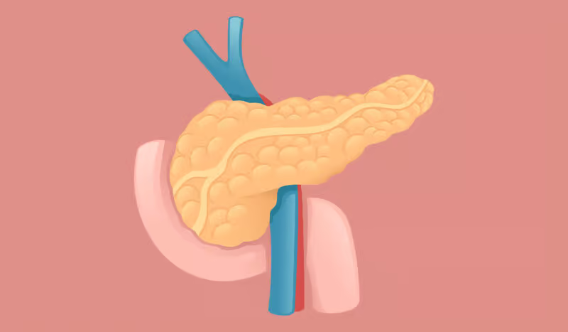
The aorta is a vital part of the human body and is affected by aortic aneurysms. Abdominal aortic aneurysms are an example of a condition that arises when the aorta in the abdomen is involved (AAA), while thoracic aortic aneurysms occur in the chest area (TAA). At times, they can be found in both areas, which is known as thoracoabdominal aneurysms.
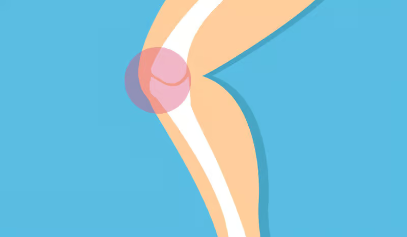
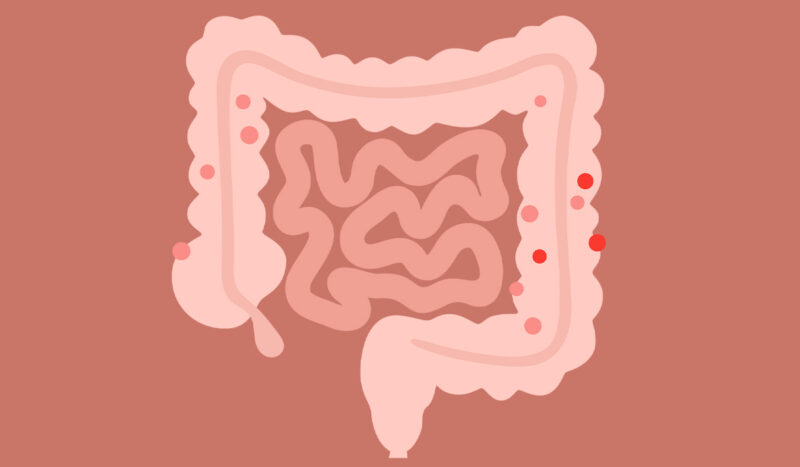
Peripheral type is the term used to describe the occurrence of them outside of the aorta or the brain, such as the blood vessels of the legs, with the popliteal artery being the most frequent site (of note is an artery situated behind the knee), the spleen, or the neck. Visceral variants are a less common form of aneurysms. They affect arteries that supply blood to the intestines or kidneys.
Table of Contents