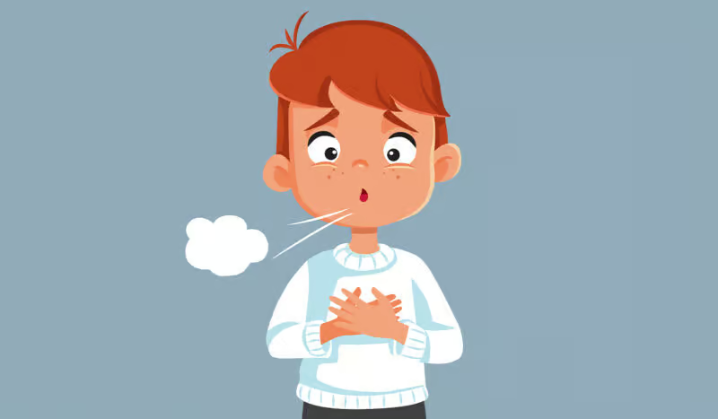
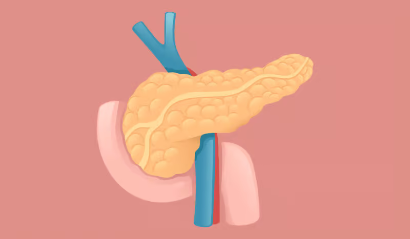
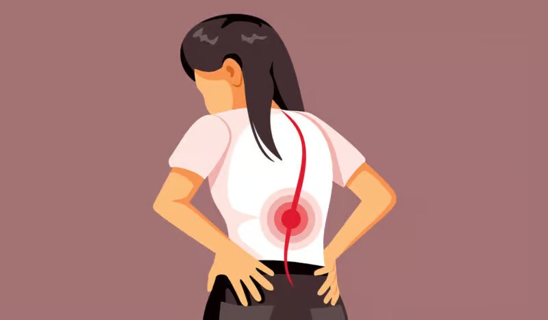
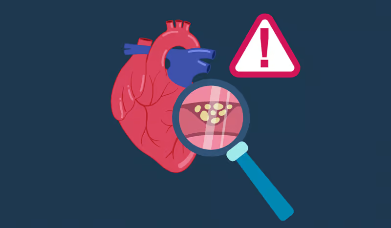
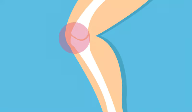
Esophagitis is simply an inflammation of the inner lining of the esophagus. Esophagus is the muscle tube through which food is transported from the mouth to the stomach. It is interesting to note that the irritation of the esophagus can also lead to the esophagus becoming red and swollen. It can be injured because of different causes.
The impact of these symptoms may be the body’s defense response. That means that only redness, slight swelling, and pain may appear. The situation gets worse and the body gets sick when the tissue is so damaged that it breaks down and ulcers or areas of erosion are formed. This can make it bleed, leave scars, or even change the esophagus’ shape and function forever.
The prevalence of esophagitis can be estimated when the population under study and the diagnostic criteria are defined. Reflux esophagitis is a common condition mostly linked with gastroesophageal reflux disease (GERD). It is that up to 20% of the adults in the West who are affected experience it at least weekly. From those, up to a significant fraction have evidence of esophageal inflammation on endoscopy.
Recordings show that almost half of the patients with the long duration of symptoms of reflux show mild to severe levels of esophagitis during examination. Nonetheless, the fact that many people are asymptomatic means the actual prevalence could be higher than these current official numbers. Unless of course, the estimates cover asymptomatic esophagitis cases as well.
The genesis of esophagitis is the result of various factors that can be categorized under the following causes. Gastroesophageal reflux disease (GERD) is the most significant cause. When stomach acid and sometimes bile are regurgitated back into the esophagus, they can generate cellular injury directly. The physical result of the exposure is the development of redness of the area, ulcerations. In the worst case, Barrett’s esophagus, a precancerous condition. The lower esophageal sphincter, a circular band of muscle located at the junction of the esophagus and stomach that functions to prevent acid reflux, can be impaired if exposed to factors like obesity, hiatal hernia, smoking, pregnancy, or some drugs.
Another cause is medication-induced esophagitis. Some medications, in particular, antibiotics such as tetracycline and doxycycline, bisphosphonates for osteoporosis, nonsteroidal anti-inflammatory drugs (NSAIDs), and potassium chloride tablets are associated with the highest risk of causing injuries to the esophagus and inducing inflammation process. The reason is that these drugs can get stuck in the esophagus. If they are not swallowed down properly, they can dissolve in the throat releasing corrosive levels. There is even more risk if a person is lying down right after swallowing the capsules without having a sufficient amount of water.
The other important cause is infections that lead to esophagitis in most people who have a weakened immune system. Predominantly; patients of HIV/AIDS, solid organ transplant recipients on immunosuppressive regimens, and patients undergoing cancer chemotherapy are the most anticipated individuals to suffer from esophagitis. The most likely causative agents are Candida, namely yeast, herpes simplex virus, and cytomegalovirus. Candida infections are identified by the presence of plaques and are visible with endoscopy while viral infections usually produce small ulcers.
Esophagitis is an umbrella term and not a single, clear-cut condition; numerous subtypes coexist, each with its idiosyncrasies. One of the most classic types is reflux esophagitis, which arises because of extensive acid exposure over time. From an obvious perspective, the organ's inner surface may get a reddish color, be eroded, or exhibit an ulcer. Histologically, the pathologist will see an increased number of basal cells, the length of the papillae increases and the neutrophils appear in the tissue in the case of infection.
Eosinophilic esophagitis is a type of esophagitis that is mainly characterized by the presence of a large number of eosinophils in the esophageal mucosa, especially on a biopsy, where the weight ratio of eosinophils to other immune cells is higher than 15/field. The major signs one will see by direct observation include falcon rings, longitudinal grooves, white residues, or a narrow lumen. It is difficult to distinguish it from reflux esophagitis. Sometimes, the only way to be sure the cause is not acid is to hold back the proton pump inhibitor therapy.
People diagnosed with esophagitis might come to the office with the widest variety of complaints. The most typical complaints are linked to dysphagia, a medical term for difficulty in swallowing. Initially, that could be the feeling of food stuck in the chest, particularly when it comes to eating solid foods.
Odynophagia, or pain with swallowing, is characteristic of ulcers and/or infections. If it doesn't occur together with reflux, then it is of common etiology. On the contrary, gastric pain caused by symptoms of reflux may show itself as a burning sensation right behind the middle of the chest bone and in the neck. Some use the term “heartburn”. Although the word itself confuses people as “chest pain” can more often point to heart conditions in the large majority of the cases. This often leads to panic attacks or necessary medical treatment to be immediately sought.
The chest pain discomfort located in the region of the retrosternum is the next most frequent appearance. The pain is quite similar to the symptoms of angina. It means it can radiate to the back, the neck, and both arms. It generally gets even worse through bending over and especially lying flat. Sour or bitter taste fluid reflux may follow these symptoms. It is especially true when the lower esophageal sphincter malfunctions, and does not close in a proper way.
The diagnostic process of esophagitis is initiated by both a complete history and a physical examination as basic modalities.
A detailed inquiry about the character and location of the chest pain, the time of the meals, any changes that may seem to make it better or worse, and any associated symptoms such as cough or hoarseness will provide helpful clues about the nature of the lesion. In addition, the history of the medication taken is important. It is because one has to know if pills may have caused the esophagitis. Also, it is necessary to ask if one has any allergies or has an atopic history. It is because this may raise suspicion of an eosinophilic cause.
Endoscopy is the process of using a thin tube with a light and a camera to look inside the human body. It helps doctors to diagnose and treat some disorders of the gastrointestinal system. Due to the fact that the esophagus begins to narrow to form a straight tube, the diameter is smaller, and the mucosa is rough; thus, the sedative only brings some comfort but without full unconsciousness. The research was carried out by scientists at the University of the South. They have used this detailed method of experiment. This method, though generally safe, with uncommon events such as bleeding or cutting while rare can occur.
The barium swallow test is a method of examining the esophagus that involves X-ray imaging. It is a standard imaging study. It is also known as an esophagram, used to diagnose diseases of the esophagus. These include gastroesophageal reflux, hiatal hernia, foreign bodies, tumors, and strictures, among others, through radiographic images or barium fluoroscopy. The barium swallow examination is useful for evaluating the esophagus, stomach, and first part of the small intestine. The patient drinks a liquid suspension of barium (that shows white on an X-ray) while being observed under the X-ray. Images are taken at different angles.
For the reason of not being completely alone, each meal must be monitored in the department of the hospital in a special room that is equipped with technological equipment to provide active visualization of the results. At the end of the observation, the results are evaluated according to the established upper limit of the time interval of the pH level registered. If such time of duration is not exceeded, the acid exposure to the esophagus is normal. The method of pH-meter, a 24-hour test, gives the opportunity to the experts to measure the acidity or alkalinity of the esophagus.
Management of esophagitis focuses on finding the cause of it, alleviating the patient's symptoms and promoting healing of the lining of the esophagus. Avoiding complications from this condition is also key. The most common tools used here are patient education and lifestyle changes, drugs, and rarely, surgery. Depending on the specific type of disease a patient has, the doctor will go for the most effective approach such as reflux, eosinophilic, infectious, or drug-induced- because universal medicine does not guarantee the recovery of the patient.
The medicines that belong to this group prevent the production of extra acid. Therefore, they are the most frequent treatment for reflux esophagitis. What they basically do is block the ATPase enzyme system at the parietal cell level in the stomach, reducing in a very significant way the secretion of gastric acid. The absorption of acid by these drugs not only prompts the mucosa to self-heal but also, provides relief of the symptoms of the patient.
Generally speaking, PPIs are taken before breakfast once a day as long as the patient needs it. It is possible that certain patients are unresponsive to this regimen. Dosage of two times a day would be their option. Nevertheless, taking them for a long time can lead to serious health conditions. These may include malabsorption of nutrients, higher chances for infection, and kidney diseases. Thus, it is best to start taking the drugs at the lowest effective dose after the patient no longer feels the symptoms of esophagitis.
Histamine H2 blocking agents such as omeprazole, lansoprazole, and esomeprazole when available, and cimetidine, famotidine, and ranitidine when available were the main drugs used at that time. These drugs interacted with histamine-2 receptors which are located on gastric parietal cells. Therefore, they were able to suppress the generation of acid in the stomach.
As far as the drugs' potency is concerned, they were not that strong. However, in a minority of mild cases, or in cases where the patient did not bear the trace elements of PPIs, the H2 blockers were effective either as drugs of choice or complementary. The usage of the drugs is twice a day but with the possibility of the night dose on the stormy night, they can tackle nocturnal acid breakthroughs. The biggest downside is tolerance which is developed over time and reduces the efficacy of the drugs over time.
Another nonpharmacological option is making a dietary change that greatly reduces the severity of the symptoms. These major causes of the condition have to be excluded from the patient's diet and life, e.g. the patient has to avoid peppermint, chocolate, caffeine, alcohol, and high-fat meals as they all together reduce the lower esophageal sphincter tone. The products that in most cases are the reason for heartburn are hot/spicy food, citrus fruits, tomatoes, and onions. If avoided, the patient will feel a lot better.
It is a well-known truth that eating fewer but frequent meals can be a good preventive measure. It is because the stomach does not distend and reflux is alleviated that way. The amount of stomach acid at night is much less and the sleep of the person is of good quality if the person lies down and their head is raised between 15 and 20 cm.
Endoscopic dilation is used in the treatment of patients with narrowings of the esophagus who still have the stricture even after medical therapy or whose rings did not give up dysphagia. This involves a gastroenterologist feeding a rubber tube that has a balloon or a specific tube through the esophagus so that endoscopic dilation can be implemented. Compression of the narrowed part happens when the balloon is slowly inflated. It may lead to the patient's feeling of discomfort experienced in swallowing.
If dilation fails to provide adequate luminal dimensions, then the narrowing part might need restretching in the future. Still, there is a limit to the amount of force that can be applied, and injury to the esophagus as a result of the procedure is a slight possibility. When performed by skillful hands, the observed negative side effects are usually mild and transient. Although the risk of mucosal tears or perforation still persists even in such circumstances.
Table of Contents